
Drs. Tom Michaud, Matt Dilnot, and Karen Mickle discuss the importance of toe strength. READ THE ARTICLE HERE.
0 Comments
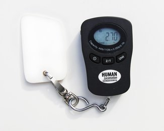 Figure 1 The ToePro Dynamometer for quick and easy measurement of foot strength In addition to providing targeted treatment for intrinsic muscles of the foot, practitioners are now able to measure strength before, during and after treatment to ensure effective therapy.
The Toe Strength Dynamometer is the first low cost and easily available instrument to assist in the measurement of foot strength in all people, including those with diabetes. The instrument evolved from the research of Professor Hylton Menz at La Trobe University in 2006 who sought to identify a simple method of measuring foot strength in a clinical setting using the age-old Paper Grip test (Menz et al., 2006). Leading author and respected biomechanics expert Dr Thomas Michaud adapted this technique to create the Toe Strength Dynamometer (Product name – ToePro Dynamometer™). Toe grip strength has been shown to detect foot muscle weakness and can predict risk of falls (Menz, Morris and Lord, 2006; Mickle et al., 2009), reduced athletic performance (Kulmala et al., 2014; Paquette, Devita and Williams, 2018), clawing of the toes (Mickle et al., 2009), and has been implicated in heel pain development (Sullivan et al., 2015; Barnes et al., 2017) and knee pain (Uritani et al., 2017) amongst other conditions. Identifying weakness and measuring response to physical therapy objectively has been difficult in a clinical setting. The Toe Strength Dynamometer gives practitioners the opportunity to solve this problem and measure physical therapy interventions for a variety of conditions in both diabetes and in a wide variety of other ailments of the lower limb. The Toe Strength Dynamometer can be used quickly and simply to create reliable, reproducible tests of foot strength and can be used as part of a standard foot assessment. With the identification of foot weakness practitioners can implement foot strength and mobilisation programs to assist in reversing the loss of strength and to maintain mobility of the forefoot (Balducci et al., 2006; Sartor et al., 2014; Kanchanasamut and Pensri, 2017). References Balducci, S. et al. (2006) ‘Exercise training can modify the natural history of diabetic peripheral neuropathy’, Journal of Diabetes and its Complications, 20(4), pp. 216–223. doi: 10.1016/j.jdiacomp.2005.07.005. Barnes, A. et al. (2017) ‘Clinical and Functional Characteristics of People With Chronic and Recent-Onset Plantar Heel Pain’, PM and R. American Academy of Physical Medicine and Rehabilitation, 9(11), pp. 1128–1134. doi: 10.1016/j.pmrj.2017.04.009. Kanchanasamut, W. and Pensri, P. (2017) ‘Effects of weight-bearing exercise on a mini-trampoline on foot mobility, plantar pressure and sensation of diabetic neuropathic feet; a preliminary study’, Diabetic Foot and Ankle. Taylor & Francis, 8(1). doi: 10.1080/2000625X.2017.1287239. Kulmala, J. et al. (2014) ‘Which muscles compromise human locomotor performance with age ? Which muscles compromise human locomotor performance with age ?’ Menz, H. B. et al. (2006) ‘Plantarflexion Strength of the Toes: Age and Gender Differences and Evaluation of a Clinical Screening Test’, Foot & Ankle International, 27(12), pp. 1103–1108. doi: 10.1177/107110070602701217. Menz, H. B., Morris, M. E. and Lord, S. R. (2006) ‘Foot and Ankle Risk Factors for Falls in Older People: A Prospective Study’, The Journals of Gerontology Series A: Biological Sciences and Medical Sciences, 61(8), pp. 866–870. doi: 10.1093/gerona/61.8.866. Mickle, K. J. et al. (2009) ‘ISB Clinical Biomechanics Award 2009. Toe weakness and deformity increase the risk of falls in older people’, Clinical Biomechanics. Elsevier Ltd, 24(10), pp. 787–791. doi: 10.1016/j.clinbiomech.2009.08.011. Paquette, M. R., Devita, P. and Williams, D. S. B. (2018) ‘Biomechanical Implications of Training Volume and Intensity in Aging Runners’, Medicine and Science in Sports and Exercise, 50(3), pp. 510–515. doi: 10.1249/MSS.0000000000001452. Sartor, C. D. et al. (2014) ‘Effects of strengthening, stretching and functional training on foot function in patients with diabetic neuropathy: Results of a randomized controlled trial’, BMC Musculoskeletal Disorders, 15(1), pp. 1–13. doi: 10.1186/1471-2474-15-137. Sullivan, J. et al. (2015) ‘Musculoskeletal and activity-related factors associated with plantar heel pain’, Foot and Ankle International, 36(1), pp. 37–45. doi: 10.1177/1071100714551021. Uritani, D. et al. (2017) ‘Relationship between toe grip strength and osteoarthritis of the knee in Japanese women: A multi-centre study’, Physiotherapy (United Kingdom), 101(25870971), pp. eS1560-eS1561. doi: http://dx.doi.org/org/10.1016/j.physio.2015.03.1559. 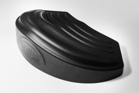 The patented ToePro strengthening device facilities the most effective and simple method of improving foot muscle strength, can be used for effective eccentric calf exercises, promotes ankle flexibility and assists in strengthening synergists of the triceps surae complex. This device paves the way for practitioners now to focus on improving ankle flexibility and promoting strengthening of the muscles of the foot. This can potentially reduce and redistribute loads away from the plantar fascia and potentially improve strength for the outside stabilizers of the ankle. These are now considered to be significant contributors to the cause of heel pain in recent research by Barnes et al. (Barnes et al., 2017) Flexor hallucis longus, a forgotten but significant muscle with a tendon a fifth the size of the Achilles, exerts force of about 50% of bodyweight during propulsion (Jacob, 2001) and will likely also improve better control of motions of the foot and ankle and reduce loads on the plantar fascia. The authors of the paper by Barnes et al from Sydney University make the following statement regarding foot strength in their discussion: “Toe flexor and ankle eversion strength was reduced in the plantar heel pain group, ……. Toe flexor weakness has been previously identified in a small sample of people with plantar heel pain, and our larger study provides further evidence that toe flexor weakness may be associated with plantar heel pain. “ Although this paper refers to only one other study with a “small sample size”, conducted by Allen and Gross in 2003, in fact there are other studies that have also identified intrinsic muscle atrophy with heel pain. This includes a study by Chang et al 2012 who found intrinsic muscle atrophy in the forefoot was associated with heel pain; Cheung et al 2016 found rearfoot intrinsic atrophy was associated with heel pain; and additionally, Wearing in 2004 also implied the involvement of foot intrinsic muscles was required to potentially offload the plantar fascia (Allen and Gross, 2003; Wearing et al., 2004; Chang, Kent-Braun and Hamill, 2012; Cheung et al., 2016). References Allen, R. H. and Gross, M. T. (2003) ‘Toe flexors strength and passive extension range of motion of the first metatarsophalangeal joint in individuals with plantar fasciitis.’, The Journal of orthopaedic and sports physical therapy, 33(8), pp. 468–78. doi: 10.2519/jospt.2003.33.8.468. Chang, R., Kent-Braun, J. A. and Hamill, J. (2012) ‘Use of MRI for volume estimation of tibialis posterior and plantar intrinsic foot muscles in healthy and chronic plantar fasciitis limbs’, Clinical Biomechanics. Elsevier Ltd, 27(5), pp. 500–505. doi: 10.1016/j.clinbiomech.2011.11.007. Cheung, R. T. H. et al. (2016) ‘Intrinsic foot muscle volume in experienced runners with and without chronic plantar fasciitis’, Journal of Science and Medicine in Sport, 19(9), pp. 713–715. doi: 10.1016/j.jsams.2015.11.004. Jacob, H. A. C. (2001) ‘Forces acting in the forefoot during normal gait - An estimate’, Clinical Biomechanics, 16(9), pp. 783–792. doi: 10.1016/S0268-0033(01)00070-5. Wearing, S. C. et al. (2004) ‘Sagittal movement of the medial longitudinal arch is unchanged in plantar fasciitis.’, Medicine and science in sports and exercise, 36(10), pp. 1761–7. doi: 10.1249/01.MSS.0000142297.10881.11. 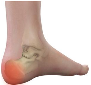 Research conducted in Sydney, Australia has clarified what has been suspected by a growing number of researchers around the world as major contributors to the cause of plantar fasciitis (heel pain). The research identified that limited ankle range, reduced foot (toe flexor) and leg strength (peroneal strength) and increased bodyweight appear to play a significant role in this condition (Barnes et al., 2017). The study conducted at Sydney University involved several leaders in musculoskeletal research. The research was significant for both the quality as well as the breadth of the research that was conducted. The study compared 71 people with long term heel pain with 61 people who had heel pain of less than 6 months. This world leading research group used this 2017 publication to extend on the research they published in 2015 in which 202 people with heel pain were compared with 70 well matched controls. Both papers evaluated an extensive range of factors (outlined below) to isolate which of these appear to be the most important in clinical assessment and treatment. The factors evaluated have at various times been implicated in the development of heel pain but required further research. The factors included: Biomechanical alignment; joint mobility; muscle strength of both the foot and leg; calf endurance; and weightbearing activity levels. Importantly this research has clarified what is not likely to be important in plantar fasciitis. Specifically heel pain was not associated with big toe range of motion (either high or low flexibility); excessive flexibility of the arch (hypermobility); calf strength or calf endurance; inwards or outwards movement of the foot; foot alignment; occupational standing time, or exercise level. Interestingly it has shined a light on the work of Rathleff et al. in 2009 which appeared to indicate that high load strength training for the calf muscles was an effective treatment for plantar fasciitis. The new research from Barnes proved this unlikely to be the case indicating both calf strength and endurance were not associated with heel pain risk. The research by Rathleff has been criticised previously. Specifically, it was criticised for its methodology which a) combined multiple treatments at once but singled out high load calf strengthening as the over-riding effective treatment b) had no control group and c) showed that strengthening took a very long time to take effect in respect to normal clinical practice i.e. 3 months and d) the groups were only different at the 3 month point and in fact the strength group were slightly, but not statistically significant, worse at the end of the study than the non-strength group (Rathleff et al., 2015). Only the non-strengthening group had subjects who reported no pain at the 12-month point. Interestingly, the natural history of plantar fasciitis even without treatment has been suggested to be 12 months (Caselli et al., 1997). 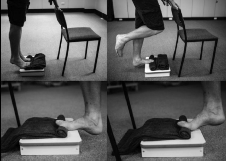 However, there are important aspects to the Rathleff study. The calf exercises in the study are done through a full range and involve eccentric loading. This is likely to be an effective way of maximising flexibility of the ankle, in keeping with the new research paper which suggested that reduced ankle range may be of significance as a cause of plantar fasciitis. Eccentric loading in a lengthened position is also likely to result in the addition of sarcomeres in series (sarcomerogenesis) to the calf muscle improving its resting length and therefore improving ankle range. These adaptations also potentially increases the joint angle at which peak torque is generated (O’Sullivan, McAuliffe and DeBurca, 2012). All of this is likely to improve better ankle control through its entire range, improve better control of forward body movement (especially in the case of increased BMI) and ultimately helps to unload the plantar fascia through the propulsive period of gait. Thus, asking patients to perform calf strengthening exercises on a step is likely to be helpful, especially through the full lower range, although we are unsure as to whether placing a towel under the toes whilst doing this is necessary. About heel pain Plantar heel pain is the most common foot disorder treated by health care practitioners (Martin et al., 2014). It occurs mainly in adults and effects people who come from both active and sedentary lifestyles. In the United States approximately 2 million people seek treatment for plantar heel pain every year, at a cost of more than $300 million (Riddle and Schappert, 2004; Tong and Furia, 2010). Heel pain is believed to occur due to excessive cumulative strain at the attachment of the plantar fascia and mechanical overload is thought to play a major role in causing this condition. Best care of heel pain has alluded practitioners for decades with researchers in the past being unable to identify the likely causative factors. References Barnes, A. et al. (2017) ‘Clinical and Functional Characteristics of People With Chronic and Recent-Onset Plantar Heel Pain’, PM and R. American Academy of Physical Medicine and Rehabilitation, 9(11), pp. 1128–1134. doi: 10.1016/j.pmrj.2017.04.009. Caselli, M. et al. (1997) ‘Evaluation of magnetic foil and PPT Insoles in the treatment of heel pain’, Journal of the American Podiatric Medical Association, 87(1), pp. 11–16. doi: 10.7547/87507315-87-1-11. Martin, R. L. et al. (2014) ‘Heel Pain—Plantar Fasciitis: Revision 2014’, Journal of Orthopaedic & Sports Physical Therapy, 44(11), pp. A1–A33. doi: 10.2519/jospt.2014.0303. O’Sullivan, K., McAuliffe, S. and DeBurca, N. (2012) ‘The effects of eccentric training on lower limb flexibility: A systematic review’, British Journal of Sports Medicine, 46(12), pp. 838–845. doi: 10.1136/bjsports-2011-090835. Rathleff, M. S. et al. (2015) ‘High-load strength training improves outcome in patients with plantar fasciitis: A randomized controlled trial with 12-month follow-up’, Scandinavian Journal of Medicine & Science in Sports, 25(3), pp. e292–e300. doi: 10.1111/sms.12313. Riddle, D. L. and Schappert, S. M. (2004) ‘Volume of Ambulatory Care Visits and Patterns of Care for Patients Diagnosed with Plantar Fasciitis: A National Study of Medical Doctors’, Foot & Ankle International, 25(5), pp. 303–310. doi: 10.1177/107110070402500505. Tong, K. B. and Furia, J. (2010) ‘Economic burden of plantar fasciitis treatment in the United States’, American Journal of Orthopedics (Chatham, Nj), 39(5), pp. 227–231.   At first glance, this study doesn't seem so surprising Fifteen men performed a heavy resistance toe flexor strength training program for their feet and ankles with 90% of their maximal voluntary isometric contraction for 7 weeks (560 contractions). At the end of the 7 week program push-off strength in the feet and ankles had more than doubled, toe strength had increased 10-15% and jump distance had increased on average about 7%.(Goldmann et al., 2013) On the face of it, it makes sense that performing high resistance training for your foot muscles would indeed be quite effective, if not a well established method of improving foot strength. That last part is where you would be wrong. It has not been a well established fact. In fact so few people considered it a possibility that almost no one has studied it. To date, there have been few attempts to measure and strengthen foot muscles while looking at changes in function. More specifically there are even fewer studies which look at the ability to specifically strengthen toe-flexor muscles and we have been somewhat sceptical that it would make the slightest difference. Further, of those that have attempted to do this before, the results have been somewhat disappointing (Spink, Fotoohabadi and Menz, 2010; Houck et al., 2017) which makes this study even more compelling. Previous studies had asked participants to exercise their muscles with loads and in positions that are not functional i.e. don't represent loads typical of those experienced when walking and in positions that don't represent how the joint is used during activities such as walking, running, jumping or sprinting. This study adds enormously to the rapidly growing research surrounding the importance of foot muscle strength. All people involved with rehabilitation, including those interested in foot, knee, hip and back function need to get with the program and grow their knowledge base. References Goldmann, J. P. et al. (2013) ‘The potential of toe flexor muscles to enhance performance’, Journal of Sports Sciences, 31(4), pp. 424–433. doi: 10.1080/02640414.2012.736627. Houck, J. et al. (2017) ‘Can Foot Exercises Alter Foot Posture, Strength, and Walking Foot Pressure Patterns in People with Severe Flat Foot?’, AOFAS Annual Meeting, pp. 1–2. doi: 10.1177/2473011417S000199. Spink, M. J., Fotoohabadi, M. R. and Menz, H. B. (2010) ‘Foot and ankle strength assessment using hand-held dynamometry: Reliability and age-related differences’, Gerontology, 56(6), pp. 525–532. doi: 10.1159/000264655. Makes clinical assessment of Diabetic foot weakness easyMelbourne, September, 12, 2018 - The way in which we assess diabetic feet in a clinical setting could change dramatically with the advent of a new device to assess muscle strength in diabetes. The ToePro Dynamometer™ is the first low cost and easily available instrument to assist in the measurement of foot strength in all people, including those with diabetes. Foot muscle weakness can develop very early in diabetes and occurs often well before sensory changes are detected in the foot. It is one of several factors which can lead to structural changes in the toes – specifically clawing and hammering of the toes. These changes are strong predictors of increased plantar forefoot pressures and together are the number one predictor of peak forefoot pressures, essentially crushing tissues under the forefoot, a major risk factor in diabetic ulcer formation. To read the full press release click here  It is surprising how long it has taken for significant steps to be made in understanding the relationship between foot muscle strength and foot function. The most recent exciting paper has come from one of the world leaders in the research of intrinsic muscles and their implications in toe deformities. Karen Mickle from Victoria University, Australia has been extraordinarily productive over the last 10 years with her use of diagnostic ultrasound and dynamometers. She has collaborated with some well known researchers too including Chris Nester and Hylton Menz. The most recent paper to have come forth from Karen has just been published in Arthritis Care & Research. Essentially the results of this paper have confirmed what has long believed to be the case i.e. if you have toe deformities such as hallux valgus and claw toes, you are likely to have atrophic (smaller and weaker) muscles. If you have hallux valgus you have a smaller abductor hallucis, if you have lesser toe deformities (claw toes) then you had atrophy and weakness in the intrinsics attached to these. 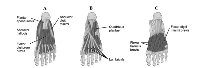 This study collected data from 44 older adults (>60 years) with either toe deformities or not (controls). They then underwent ultrasound assessment to look at the size of the muscles in their feet with a standardized protocol. What was particularly interesting in this study was that virtually every muscle examined (of which there were 7 different muscles examined) was atrophic relative to the controls, bar flexor hallucis longus. Take a look at the chart below: 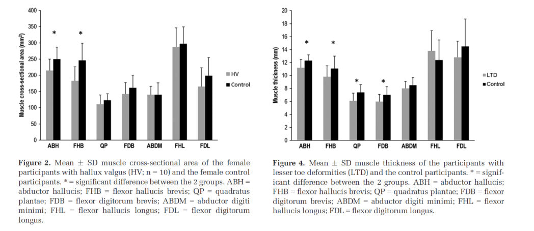 The tricky part of this research then leads us to the next important question. Does the atrophy occur because of the toe deformity or does it lead to the toe deformity? Current evidence would point to the latter, which suggests specifically in relation to hallux valgus that prevention through exercise should happen in the early stages to stop the progression of the deformity. Once the abductor hallucis muscle moves under the 1st metatarsal it acts as a weak flexor instead of an abductor.
References:
This interview with Dr Tom Michaud was conducted on 19th December 2017 with the "2 Movement Guys", Adam Wolf,PT and Nicholas Studholme, DC. It is an enjoyable general chat about orthoses, strength and some general lower limb management tips. "Interested in orthotics, biomechanics, and foot function? Check out the recent podcast with Dr. Tom Michaud D.C., author of Human Locomotion: The Conservative Management of Gait-Related Disorders, as used in physical therapy, chiropractic, pedorthic, and podiatry schools around the world. Tom is a wealth of clinically relevant and easily applicable information. This is going to be worth your time!" Podcast here: References in this Podcast include: Titanium rods and tibio-femoral rotation
Samuel Adams Beer - Boston Lager |
AuthorMatt Dilnot is a Podiatrist working in the Eastern Suburbs of Melbourne, Victoria. Archives
May 2020
Categories
All

|
 RSS Feed